When you visit PVSC for a glaucoma evaluation, you’ll go through some comprehensive eye exams and tests to allow us to efficiently get the most accurate information about your eyes so that you make the least number of visits to the office. Your eye health is important to us as well as your time.
Some of these tests are covered by OHIP but some are not. Insurance companies and Ontario Disability Support Program (ODSP)/Ontario Works also cover some of these tests. You’ll be informed of the cost of any testing that you would need beforehand and you have the right to accept or refuse any test that involves an extra cost.
More important than the results of these tests are the changes that happen overtime. If the test results are stable, the glaucoma is well-controlled. However, if the results are progressive, the glaucoma isn’t well-controlled. If you have abnormalities that aren’t changing over time, it’s possibly because they’re not caused by glaucoma as these changes would be progressive.
One of the problems with glaucoma is that there are typically no symptoms in the early stages. Many people who have the disease don’t know they have it which is why it’s important to have regular eye exams, especially as you get older. Here are some of the tests you can expect from a glaucoma evaluation with Dr. Youssef:
Your pupils will be dilated (widened) and numbed with eye drops to allow us a better view of the inside of your eye.
We measure the pressure inside of your eye, an important part of glaucoma evaluation because glaucoma can be caused by high pressure. Healthy eye pressure generally ranges between 10-21 and a high pressure reading is often the first sign that you have glaucoma.
The drainage angle is the area between the iris and the cornea and it’s where the aqueous humor drains out. Sometimes the drainage angle is blocked or simply isn’t working as it should so the liquid can’t escape and causes the pressure inside the eye to rise. During a gonioscopy exam, Dr. Youssef uses a mirrored lens to check the drainage angle and determine the type of glaucoma you have.
Your visual field is how wide your eye can see when you’re focused on a single point. By testing how well you see objects in your visual field, Dr. Youssef can determine how much vision has been lost over time, a crucial part of a glaucoma diagnosis to determine if it’s stable or getting worse. A visual field test can also show if and where blind spots appear in your peripheral vision, including spots you might not even notice.
The test is performed using a bowl-shaped machine called a perimeter. When taking the test, a patch covers one of your eyes so that only one eye is tested at a time. Random points of light will flash around the bowl-shaped perimeter and you’ll be asked to press a button whenever you see a light.
Thickness of the cornea can affect eye pressure readings, so a pachymeter is gently placed on the cornea to measure its thickness. However, at PVSC, we prefer to measure corneal thickness using optical coherence tomography (OCT), because it’s more accurate, non-touch, and offers us much more information than pachymeters.
Optical coherence tomography (OCT) testing has become the standard of care for diagnosis and treatment of many ocular diseases. OCT is a non-invasive tool which uses harmless light waves to take high-resolution, detailed images of the back and front sections of the eye. This cutting-edge technology can produce live, cross-sectional scans of the front of the eye to allow us to analyze the cornea, the anterior chamber angle, the iris and lens in excellent quality. These scans allow us to directly observe the drainage angle so we can easily identify if glaucoma is caused by a blockage.
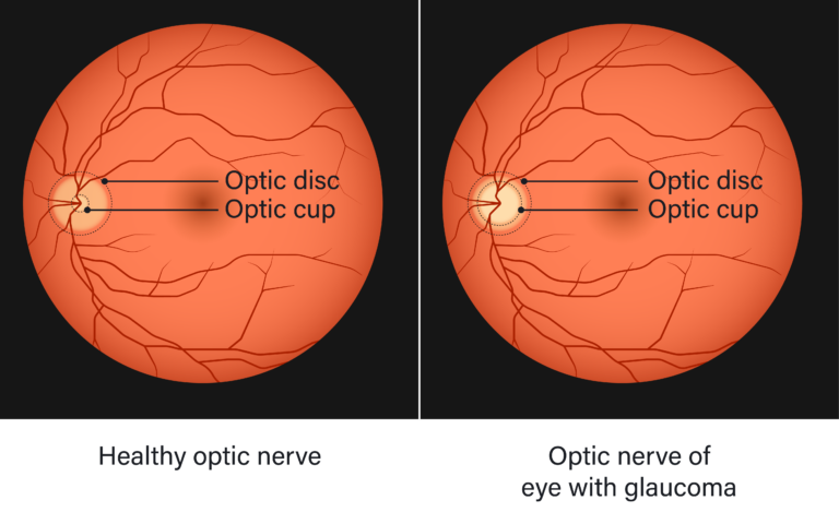
A healthy optic nerve is made up of more than one million tiny nerves. Glaucoma is the result of damages to these nerves which changes its appearance. Dr. Youssef may use an ophthalmoscope to magnify the inside of the eye and see the back of your eye to inspect your retina and optic nerve for signs of damage.
Fundus photography also takes photos of the interior surface of the eye which help monitor progression or diagnosis of a disease. Compared to an ophthalmoscope, fundus photographs generally capture much larger areas of the interior of the eye than what can be seen at any one time with ophthalmoscopes (by more than 80%!). To monitor glaucoma, fundus photos allow us to observe the sizes of the optic cup and optic disk as shown in the diagram below. Increases in the size of the optic cup compared to the optic disc can tell us damage is still occurring to the nerves.