
Please see a physician or visit the emergency room if you have any sudden changes in your vision. If they suspect or conclude you have a retinal tear or retinal detachment, you will likely be urgently referred to Dr. Youssef.
These are the common tests and examinations that you’re required to have when you come for a consultation or a follow-up for a retinal disease. These tests are arranged in order to achieve the most accurate information about your eyes in the most efficient way. Your eye health as well as your time is important to us.
It’s important to note that some of these tests are covered by OHIP while others are not. You’ll be informed of the cost of any testing that you would need before it’s done and you have the right to accept or refuse the tests that involve an extra cost. Insurance companies and ODSP/Ontario works to cover some of these tests.
Because there’s often a lot of ground to cover at your appointment, it’s a good idea to be well prepared.
Please bring your health card, any eye drops you use, a list of current medications, and your eyeglasses and sunglasses. You’ll be asked for them upon arrival.
Arrange for a driver. You won’t be able to drive the day of your appointment because the dilating drops will give you hazy/blurry vision for a few hours.
Bring a friend or family member if you’d like! Although it’s not a requirement, bringing someone you trust with you is a good idea because we’ll discuss a lot of information so extra support is always nice.
Please be aware that some of these tests are covered by OHIP but some are not. You’ll be informed of the cost of any testing that you would need before it is done and you have the right to accept or refuse these test that involve extra cost. Insurance companies and ODSP/Ontario Works cover some of these tests.
Before you begin your diagnostic testing, you’ll be asked some basic questions to give us some insight into your condition which will allow Dr. Youssef to provide you a more accurate diagnosis. You can prepare by writing down any symptoms you’re experiencing including even those that may seem unrelated and key personal information (major stresses or life changes).
At PVSC, we conduct a series of diagnostic tests so we have a comprehensive understanding of your retinal issue and figure out an effective treatment plan. During your assessment, a technician will sit with you to take your medical history and address your visual symptoms and concerns. Then they will perform the following preliminary tests:
We will measure the pressure inside your eye.
Your pupils are dilated (widened) by medicated drops on your eye. This allows us to look inside your eye through a magnifying glass to detect signs of damage or other eye issues.
This is an eye chart test to determine your ability to see clearly at different distances.
After these tests are complete, you will see Dr. Youssef who will perform a thorough examination of your eyes. Based on your test results and examination, if he suspects you have a retinal disorder, he might request more testing to confirm his diagnosis. Here are some of the tests you can expect from a retinal assessment with Dr. Youssef:
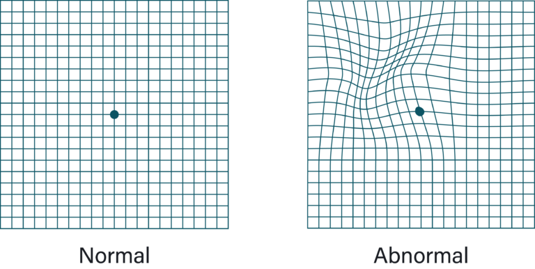
Using what’s called an Amsler grid (a grid with horizontal and vertical lines and a dot in the middle), you’ll be asked to cover each eye one at a time and stare at the black dot in the center. If the straight lines appear broken, crooked, wavy, bent, or distorted, this may indicate a retinal condition.
An ophthalmoscope is an instrument with a bright light and a special lens to examine the inside of your eyes. It shows a highly detailed 3D view, allowing the doctor to see any retinal holes, tears or detachments.
A slit-lamp is a type of microscope used to examine the front part of the eye, including the eyelids, conjunctiva, sclera, cornea, iris, anterior chamber, lens, and also parts of the retina and optic nerve.
Fundus photos accurately document the changes that happen with age and the progression of retinal conditions.
Optical coherence tomography (OCT) is a non-invasive scanning laser that provides high-resolution images of the retina. This scan measures the thickness of the retina which gives us invaluable information about its thickness and can detect any fluid buildup behind the retina so we can determine severity of any macular edema (swelling).
Fluorescein angiography is a procedure that uses a special camera to take a series of photographs of the retina after a small amount of yellow dye (fluorescein) is injected into a vein in your arm. Photos of retinal vessels can show which blood vessels are leaking fluid and how much is leaking among other things.
If Dr. Youssef can’t see the retina due to blood clouding the view, an ultrasound test may be done in the clinic. An ultrasound uses sound waves which travel through the eye and bounce off structures to create an image of the back of the eye. Using this, we can “see” through the blood to determine if your retina has detached.